
Images in Radiology
Worsening thigh pain after blunt trauma
LT Kendall Lane MD MC USN
A 19 year-old otherwise healthy male presented with right thigh pain for three weeks after another player's knee struck him playing basketball. During the first week following the injury, he noted significant improvement in the pain with NSAIDs and ice. However after the initial improvement and return to activity, the right thigh pain gradually worsened and inhibited his ability to lift his leg and walk up stairs. The patient denied any weight loss, night sweats, fever, or family history of malignancy. On physical exam, tenderness was elicited over the right quadricep and a faint resolving ecchymosis was noted. A mass was not palpable. His ability to extend his right knee was mildly reduced, and he experienced significant pain with knee flexion.
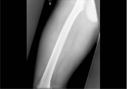
Plain radiographs were ordered because of worsening pain and unresponsiveness to conservative treatment with NSAIDs and ice. The x-ray showed peripheral soft tissue calcification consistent with myositis ossificans.
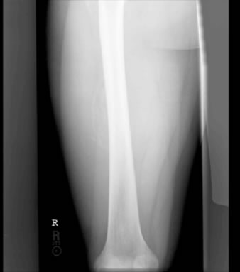
The patient was treated with rest, NSAIDS, and gentle stretching. His pain improved over the course of several weeks, and he returned to playing basketball without any difficulty. Follow-up radiographs showed progressive central calcification consistent with an evolving myositis ossificans. The cortex of the femur remained intact without focal defect.
Myositis ossificans is a benign heterotopic ossification of soft tissue characteristically associated with direct trauma. Athletes, more specifically those participating in contact sports, are at risk for developing myositis ossificans as a complication of a contusion. The lesion typically occurs within the extremities 1-3 but has been reported in other areas of soft tissue and muscle such as the chest wall4.
Calcification within the lesion of myositis ossificans will appear radiographically in the third to sixth week after injury5. A radiodense area of mature bone is seen on the periphery of the lesion. The center of the lesion is less radiodense without the presence of mature bone.
The clinician must consider osteogenic sarcoma prior to making a definitive diagnosis of myositis ossificans. There are several clues that distinguish these two very different pathologies. Plain radiographs can provide some insight. In contrast to the peripheral rimming seen in myositis ossificans, calcification typically extends from the center to the periphery in osteogenic sarcoma. The location of the lesion can also assist in making the correct diagnosis. Myositis ossificans typically develops over the diaphysis rather than the metaphysis as seen in the majority of cases of osteogenic sarcoma. Other helpful hints associated with myositis ossificans include improvement in pain over time with rest and an intact cortex on plain radiographs. The pain associated with sarcomas worsens with time and the cortex is often violated6. Histologically the two diagnoses also differ. Biopsies of myositis ossificans reveals a zone phenomenon consisting of a central undifferentiated fibrovascular zone, an osteoid formation zone, and an outer peripheral mature lamellar bone zone1-3, 7.
These histological zones can be appreciated ten days after the injury. If a biopsy is preformed prior to ten days or if a biopsy is taken from the central zone of the lesion, the pathology may resemble osteosarcoma. If there is any doubt in the diagnosis with plain
radiographs, A CT, MRI, and/or a biopsy of the lesion should be pursued.
Patients with myositis ossificans should not continue to play sports or use the affected muscle. Heat and massage should be avoided. Reinjury to the same area, returning to activity too early, or initial passive forceful stretching can lengthen recovery. Treatment includes NSAIDS, rest, and gentle stretching. Surgery may be indicated if the patent experiences persistent pain, but should not be pursued prior to a year following the initial injury.
References
1. Harmon D. Case 38, 1994. N Engl J Med 1994;331:1079-1084.
2. Resnick D. Soft tissues. In: Diagnosis of Bone and Joint Disorders. Resnick D. 3rd edn. Philadelphia, W.B. Saunders Co., 1995; pp. 4577-4584.
3. Nuovo M, Norman A, Chumas J, Ackerman L. Myositis ossificans with atypical clinical, radiographic, or pathological findings. Skel Radiol 1992;21:87-101.
4. Nisolle J-F, Delaunois L, Trigaux J.P. Myositis ossificans of the chest wall. Eur Respir J 1996;9:178-179.
5. Fu FH, Stone DA. Sports injuries; mechanisms, prevention, treatment. Philadelphia: Williams and Wilkins, 1994:758-9.
6. Operative Orthopaedics. Cambell. 9th edition, volume one1998 Mosby-Year Book, Inc. 758-759.
7. Weiss SW, Goldblum JR: Myositis ossificans. In: Enzinger and Weiss's Soft Tissue Tumors. St Louis, Mosby, 2001, Ed 4, pp 1389-1397.
First Published April 2006
Home • Journals • Search • Rules for Authors • Submit a Paper • Sponsor us
All pages copyright ©Priory Lodge Education Ltd 1994-

