Browse through our Journals...
Monocephalus Diprosopus (Complete Craniofacial Duplication) Associated with Hydrancephaly and Other Congenital Anomalies.
Dhaliwal, Adinkra, Ennis, Green
History and Presentation
A 22 year old in her third pregnancy, booked in the antenatal clinic at 10 weeks gestation. She had a base line scan which confirmed the gestational age at 10 weeks and four days. The heartbeat was visualised with normal appearances.
Her previous pregnancies were complicated by anaemia. Labour was induced for both the pregnancies due to post-maturity. There was no complications- intrapartum or postpartum. Birth weights were 4,018 and 3,656grams respectively, with no abnormalities detected.
Routine serum maternal infection screening in the current pregnancy for: Syphilis, Rubella, Hepatitis B surface antigen and Human Immunodeficiency virus were unremarkable. The patient declined serum screening for Downs’s syndrome and Spina bifida.
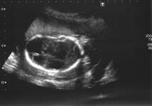
Fig 1- Hydranencephaly.
She then attended a routine anatomy scan at 20 weeks gestation and was found to have the following – oligohydramnios, abnormal appearances of the kidneys, spine and head. The lateral ventricles of the brain were dilated and the head was lemon shaped. The cerebellum was not seen. Both the kidneys were grossly enlarged and multi cystic. There appeared to be a spinal abnormality. The patient was then referred to a fetomaternal specialist for a second opinion. The findings were of conjoined heads with two sets of eyes and both brains were hydrancephalic with absence of cerebellar tissue (Fig1 &2). Left diaphragmatic hernia, short barrel-shaped chest, bilateral multicystic kidneys (Fig3), rocker- bottom type feet, abdominal distension and abnormal spinal structure.
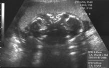 Fig.2 Conjoined Heads
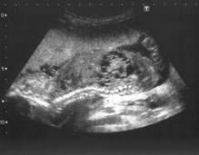 . Fig 3-Multicystic dysplasia of the kidneys and Kyphosis and scoliosis of thoracic and lumbar spine.
The parents were counselled that the multiple foetal anomalies was likely to represent a syndrome or an aneuploidy. The risk of obstructed labour was also explained due to enlarged head and abdominal circumference. The couple opted initially for further invasive assessment. Foetal blood sampling was performed and was sent for cytogenetical analysis. The received sample was inconclusive as it was of maternal origin. Due to the complex abnormality, the couple were further counselled about Termination of the pregnancy and obtaining a full post mortem examination in order to elucidate the full extent of the anomaly.
Fetocide was carried out at 21 weeks with potassium chloride (KCL) intracardiac injection. Once, fetal cardiac arrest was confirmed, the intracerebral fluid was removed in order to decompress the head(s). Medical management for termination was then commenced.
Findings at post mortem:
X-Ray and MRI confirmed two fetal faces and skull bases. Single cervical spine with multiple thoracic vertebral segmentation anomalies with no other major skeletal abnormality. Bone age was consistent with 21 fetal weeks.
General external and internal examination concluded: A male conjoined symmetrical twin of “Y” type (Terata catydidymus) with a single lower body, two faces, one head (Disprosopus) and normal upper and lower extremities (Fig 4).
Fig4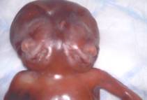

There was a left sided diaphragmatic hernia, secondary lung hypoplasia, congenital heart disease (VSD, overriding aorta and hypo plastic ascending, descending aorta and aortic arch), bilateral dysplastic cystic kidneys, hypoplasia of urerters and urinary bladder, spina bifida aperta of thoracic and lumbar segments and two, non fused brains, each with two hemispheres of various size (two pairs of olfactory and optic nerves and two hypophyses), absent cerebellum and a hypoplastic midbrain. Beneath the scalp were two entirely separate frontal bones and two partly fused parietal bones that were facing the anterior aspect of the head. The posterior aspect of the head consisted of two parietal and a partly fused single bi-lobed occipital bone. The trunk was extremely short and broadened with a shortened barrel-shaped chest. There were two pairs of eyes, two separate noses and mouths. The mandibulae were separate but hypotrophic and under-developed. There were two separate tongues that were fused to their roots creating one broader pharynx with a normal-looking epiglottis, larynx, and single trachea showing a normal bifurcation for the two main bronchi. There was a single heart with a normally positioned liver, spleen and pancreas. The gastrointestinal tract and mesentery were also normally rotated. Cytogenesis on cord and placental tissue samples did not detect any chromosomal abnormality. A normal male karyotype was recorded. On further genetic studies there was no evidence of isochromosome 12p, which is associated with diaphragmatic hernia. There was also no histological evidence of cerebellar tissue.
Discussion
Conjoined twin’s results from an embryological disturbance in the separation of twins during the 2nd week of pregnancy (12-13 days) as a result of abnormal splitting of the post – implantation blastocyte. Such incomplete separated germinal discs lead to this extremely rare fetal anomaly. The reported incidence is 1:50,000 to 1:200,000 births and 1% of monochorionic twins.
They are classified in three groups: 1) Terata Catydidymus – Diprosopus, Dicephalus, Ischiopagus (6-20%) and pyopagus (10-20%). 2) Terata Anadidyma – Dipygus, Syncephalus and Crainopagus (6-12%). 3) Terata Anacatadidyma – Thoracopagus (30-40%), Omphalopagus (25-30%) and Rachipagus. The majority of conjoined twins are thoracopagus and omphalopagus – approximately 75%. Monocephalic diprosopus conjoined twins are rare form characterized by a single body, one unusual head, two faces or a spectrum of duplication of craniofacial structures. Diprosopus results in an error related to the neurulation of the embryo (Moore KL. 1988). Complete craniofacial duplication, as in this case, may be due to the forking or bifurcation of the notochord rostrally, with the formation of two side-by-side vertebral axes, neural plates and their neural crest derivatives (Machin G, 1993).
In most of the reported cases the foetuses were anencephalic, where duplication of the brain was impossible (Gorin et al., 1990). In this particular case, there were two separate brains each showing two hemispheres, two pairs of olfactory and optic nerves, two hypophyses and one midbrain with no detectable cerebellum.
There is a high incidence of malformations that are unrelated to the point of conjunction. These include: Neural tube defects, diaphragmatic hernia, cleft lip and palate (Chervenak et al., 1985) and imperforate anus. Other associated anomalies are: Anencephaly, spina bifida, encephalocele, holoprosencephaly, hydrocephaly, abnormalities of the cranial nerves (Barr M. 1982), tetralogy of Fallot, membranous ventricular septal defect, atrial septal defects, artioventricular canal malformation and dextrocardia, double outlet right ventricle and patent foramen ovale (Turpin et al., 1981). The female: male ratio is 2:1 in up to 90-95% of the reported cases. A normal chromosomal analysis has been reported by various authors (Moerman et al., 1983; Sharony et al., 1993; Rai et al., 1998 and Wu et al., 2002). In this case, molecular cytogenesis did not detect any chromosomal abnormality.
In conclusion, we present a case report of a foetus with complete facial duplication that had intact calvaria with multiple anomalies.
References
1) Barr M. 1982. Facial duplication: case, review, and embryogenesis. Teratology 25:153-159
2) Chervenak, F.A., Pinto, M. M., Heller, C.I., Norooz, H. (1985). Obstetric significance of foetal crainofacial duplication. A case report, J. Reprod. Med., 30, 74-76.
3) Gorlin, RJ, Cohen, MM Jr, Levin, LS, editors. 1990. Syndromes of the head and neck, 3rd ed. New York: Oxford University Press.
4) Machin, G. 1993. Conjoined twins: implications for blastogenesis. Birth Defects Orig Artic Serv 29: 141-179.
5) Moerman, PH., Fryns, JP. Goddeeris P, et al 1983. Aberrant twinning (Diprosopus) associated with anencephaly. Clin Genetics 24:252-256.
6) Moore, KL 1988. The developing human: clinically oriented embryology, 4th ed. Philadelphia: W.B. Saunders Company.p.53-59.
7) Sharony R, et al. 1993. Diprosopus: A pregastrulation defect involving the head, neural tube, heart and diaphragm. Birth defects Orig Serv 29:201-209.
8) Rai VS, et al. 1998. Antenatal diagnosis of complete facial duplication: A case report of a rare craniofacial defect. Prenatal Diagnosis 18:618-620.
9) Turpin, IM, Furnas, DW, Amlie, RN. 1981. Craniofacial duplication (Diprosopus). Plast Reconstruct Surgery 67:139-142.
10) Wu, J., Staffenberg, DA. Mulliken, JB., and Shansken, AL. Diprosopus: A unique case and review of the Literature. Teratology 66:282-287 (2002).
About the authors:
Harjit Dhaliwal, M.B.B.S, D.F.F.P, Paul Adinkra M.B.B.S, Diane Ennis, RGN, RM, PGC, Pauline Green MRCOG.Drs Dhaliwal and Adinkra are Specialist Registrars, Diane Ennis is a midwife sonographer and Pauline Green is a Consultant Fetomaternal specialist all based at Arrowe Park Hospital , Wirral, United Kingdom.
E-mail – harjitdhaliwal2004@yahoo.co.uk
Copyright Priory Lodge Education Limited 2007
Firts Published November 2007
Click
on these links to visit our Journals:
Psychiatry
On-Line
Dentistry On-Line | Vet
On-Line | Chest Medicine
On-Line
GP
On-Line | Pharmacy
On-Line | Anaesthesia
On-Line | Medicine
On-Line
Family Medical
Practice On-Line
Home • Journals • Search • Rules for Authors • Submit a Paper • Sponsor us
All pages in this site copyright ©Priory Lodge Education Ltd 1994-


