Consequences of Severe Acute Generalized Exanthematous Pustulosis (AGEP) Necessitating Burn Unit Admission
Christopher R. Newey, D.O., M.S., Cleveland Clinic, Neurological Institute, Department of Neurology, Cleveland, Ohio
Aarti Sarwal, M.D., Cleveland Clinic, Neurological Institute, Department of Cerebrovascular Disease, Cleveland, Ohio
Priyanka Sharma, M.D., Cleveland Clinic, Department of Medicine, Cleveland, Ohio
Deborah Tepper, M.D., Cleveland Clinic, Neurological Institute, Department of Headache, Cleveland, Ohio
Key Words: AGEP, hyponatremia, sepsis, burn unit, hypothermia
The work was performed at Cleveland Clinic in Cleveland, Ohio.
ABSTRACT:
Acute generalized exanthematous pustulosis (AGEP) is a rare complication associated with medications, particularly antibiotics. The classic description is an eruption of pustules, sometimes confluent, resulting in Nikolsky sign, where light rubbing of the skin results in exfoliation of the outermost layer. It is typically reversible once the offending agent is stopped. We present a case of a 51-year-old-male with exfoliative dermatitis, after treatment with cefazolin and ampicillin/sulbactam diagnosed on punch biopsy as AGEP. He developed complications of hypothermia, hypernatremia, and respiratory failure requiring admission to burn unit. This case highlights a severe clinical spectrum of AGEP caused by antibiotics requiring a prompt change in medication and prudent monitoring of volume status and metabolic function. Severe cases, such as this patient, require admission to a burn unit. A review of AGEP follows this case report.
CASE
A 51-year-old-male with a history of hypertension, alcohol abuse, and non-ischemic alcoholic cardiomyopathy, presented to an outside hospital, where he was treated for anasarca and malnutrition. During this hospitalization, he developed ophthalmilitis with nasal methicillin resistant staphylococcus aureus (MRSA) and coagulase negative staphylococcus aureus (CoNS) of the right eye, and urinary tract infection with Carbapenem Resistant Acinetobacter (CRAB). He was treated with cefazolin for two days, but developed a nonspecific dermatitis of the abdomen, face, extremities, and groin. Cefazolin was changed to ampicillin/sulbactam. The rash progressed to a generalized exfoliative dermatitis and his mental status deteriorated. The patient was then transferred to our hospital. He was noted to be afebrile, hemodynamically stable, confused, hyperreflexic with bilateral lower extremity clonus. He had prominent jugular venous distention with a 3/6 apical, systolic ejection murmur and intact peripheral pulses.
Skin examination was significant for desquamation of the face, extremities, and chest sparing the groin and back (Figures 1A and 1B). The desquamation was confluent with a Nikolsky sign i.e., light rubbing of the skin resulted in exfoliation of the outermost layer. Laboratory work up was significant for a sodium of 153 mEq/L, BUN of 55 mg/dL , creatinie of 2.04 mg/dL, albumin of 2.0 g/dL.. and white cell count of 20.5k cells/mL. Chest x-ray showed an infiltrate in left lung base.
His antibiotics were changed to linezolid and tigecycline. Given the prominent skin desquamation, oral mucosal sloughing was a concern, but a laryngoscopy was unremarkable. A skin punch biopsy was performed. The pathology was interpreted as acute generalized exanthematous pustulosis.
Over the next twenty-four hours, his respiratory and mental status continued to deteriorate, and his hypernatremia worsened, requiring medical intensive care unit (MICU) admission. His metabolic abnormalities resolved with aggressive hydration and wound care with aquaphor to the whole body twice daily and silver sulfadiazine to the lower extremities. Once the rash improved, he was discharged home for outpatient follow up but was subsequently re-hospitalized with sepsis caused by skin infection. His blood cultures grew CRAB, MRSA, and candida albicans. His mental condition and rash continued to worsened, and he was transferred to a burn unit where he eventually expired.
This case highlights a rare, but life-threatening form of AGEP resulting from antibiotics. The patient developed AGEP with severe desquamation resulting in hypothermia and hypernatremia from fluid loss. The desquamation provided a nidus for bacterial growth, eventually requiring burn unit care.
DISCUSSION and REVIEW OF AGEP
AGEP is a rare condition resulting in erythematous, edematous eruptions of numerous sterile pustules, and fever with or without serum leukocytosis.1, 2 After the initial eruption, skin desquamation occurs.1 It was first described and termed by Baker and Ryan in 1968 after reviewing 104 cases of pustular psoriasis.3 But it was Beylot et al. in 1980 who termed the drug eruption pustuloses exanthématique aiguës généralizés (PEAG), which is translated AGEP, after reviewing four cases that were also initially felt to be psoriatic.4 Since then, the skin eruption has been widely reported in the literature in all age groups.5
AGEP is most commonly observed following antibiotic treatment, particularly with beta-lactams, cephalosporins, quinolones, or macrolides.1, 6-8 Non antibiotic causes, including diltiazem, mercury, thalidomide, and the antifungal terbinafine, have been described.1, 6-9 The characteristic drug eruption usually begins as pruritus followed by pinhead-sized, nonfollicular sterile pustules one to two days post drug exposure.10, 11 It commonly begins on the face, hands, and intertriginous areas with eventual mucosal involvement in 20% of cases.6, 11 If a confluence of pustules occurs, a positive Nikolsky sign may be identified.6 The rash usually resolves within two weeks with sequalae of generalized skin desquamation.10, 11 Without treatment, the mortality reaches five percent.1 Thus, it is important for the clinician to identify AGEP, but also differentiate it from other skin eruptions.
The differential for AGEP includes acne or folliculitis, but also comprises more disseminated eruptions such as acute generalized pustular psoriasis (AGPP), Sneddon-Wilkinson disease, pustular vasculitis, drug rash with eosinophilia and systemic symptoms (DRESS), toxic epidermal necrolysis (TEN), and Stevens-Johnson syndrome. The diagnosis of AGEP begins with a careful history, noting the timing of eruptions and any associated medications, particularly those described in AGEP. Patch testing may be used to rule out AGEP, but the sensitivity is only about 50%.12 The benefit of patch testing is the quick turnaround for results, usually within six hours, and the ease of performing it in less monitored settings.13 Additional testing includes the mast cell degranulation (MCD) test and the macrophage migration inhibition factor (MIF).14 A skin biopsy is the gold standard.14 The histopathology from a biopsy characteristically shows spongiform pustules under the stratum corneum.1 The biopsy may also show papillary dermal edema and perivascular polymorphous infiltrate (neutrophilic) as well as leukocytoclastic vasculitis and focal necrotic keratinocytes.1 The mechanism of AGEP is largely unknown. It is likely an immunologic phenomenon dependent upon memory T cells producing IL-3 and IL-8.6, 15 This would explain why some medications cause a delayed eruption, while others produce a more immediate reaction, presumably the result of a “re-challenge”.7
Once AGEP is suspected, the treatment includes stopping the offending agent. However, the course of AGEP inevitably continues for 8-10 days after the initial insult. Since a majority of patients have an underlying infection, a change in antibiotic is usually necessary. Additional treatment options may include topical steroids and symptomatic treatment.1 If wide-spread, severe skin desquamation occurs, as in our case, treatment for second degree burns is appropriate, with careful monitoring of volume status and other complications from extensive skin desquamation.16
CONCLUSION:
This case illustrates the importance of rapid and accurate diagnosis of AGEP. Our patient initially developed the lesions while receiving cefazolin. Once identified, the therapy was changed to ampicillin/sulbactam. However, the extensive skin desquamation provided a nidus for polymicrobial infection, requiring ongoing modification of his antibiotic treatment. The desquamation also resulted in dangerous insensible water loss resulting in recurrent, symptomatic hypernatremia necessitating burn unit admission. It is essential to recognize early AGEP and its complications, in order to effectively treat the disorder and its complications. While changing or stopping the antibiotic is essential, patients will still go through the phases of the disorder, including the desquamation phase.
FIGURE 1:
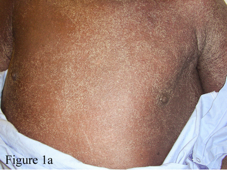
A: Thorax and abdomen of a 51-year-old male with acute generalized exanthematous pustulosis (AGEP) with pinhead-sized pustules with confluence and desquamation of the skin.
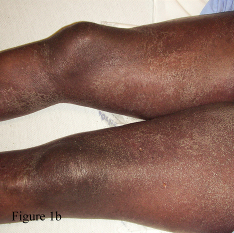 B: Lower extremities of the same patient with AGEP. Again seen are the confluence of pustules and desquamation of the skin.
REFERENCES:
1. Roujeau JC. Clinical heterogeneity of drug hypersensitivity. Toxicology. Apr 15 2005;209(2):123-129.
2. Knowles SR, Shear NH. Recognition and management of severe cutaneous drug reactions. Dermatol Clin. Apr 2007;25(2):245-253, viii.
3. Baker H, Ryan TJ. Generalized pustular psoriasis. A clinical and epidemiological study of 104 cases. Br J Dermatol. Dec 1968;80(12):771-793.
4. Beylot C, Bioulac P, Doutre MS. [Acute generalized exanthematic pustuloses (four cases) (author's transl)]. Ann Dermatol Venereol. Jan-Feb 1980;107(1-2):37-48.
5. Meadows KP, Egan CA, Vanderhooft S. Acute generalized exanthematous pustulosis (AGEP), an uncommon condition in children: case report and review of the literature. Pediatr Dermatol. Sep-Oct 2000;17(5):399-402.
6. Sidoroff A, Halevy S, Bavinck JN, Vaillant L, Roujeau JC. Acute generalized exanthematous pustulosis (AGEP)--a clinical reaction pattern. J Cutan Pathol. Mar 2001;28(3):113-119.
7. Sidoroff A, Dunant A, Viboud C, et al. Risk factors for acute generalized exanthematous pustulosis (AGEP)-results of a multinational case-control study (EuroSCAR). Br J Dermatol. Nov 2007;157(5):989-996.
8. Talati S, Lala M, Kapupara H, Thet Z. Acute generalized exanthematous pustulosis: a rare clinical entity with use of piperacillin/tazobactam. Am J Ther. Nov-Dec 2009;16(6):591-592.
9. Taberner R, Puig L, Gilaberte M, Alomar A. Acute generalized exanthematous pustulosis induced by terbinafine. Eur J Dermatol. May-Jun 2003;13(3):313-314.
10. Roujeau JC, Bioulac-Sage P, Bourseau C, et al. Acute generalized exanthematous pustulosis. Analysis of 63 cases. Arch Dermatol. Sep 1991;127(9):1333-1338.
11. Wolff K, Johnson RA, Fitzpatrick TB. Fitzpatrick's color atlas and synopsis of clinical dermatology. 6th ed. New York: McGraw-Hill Medical; 2009.
12. Wolkenstein P, Chosidow O, Flechet ML, et al. Patch testing in severe cutaneous adverse drug reactions, including Stevens-Johnson syndrome and toxic epidermal necrolysis. Contact Dermatitis. Oct 1996;35(4):234-236.
13. Beylot C, Doutre MS, Beylot-Barry M. Acute generalized exanthematous pustulosis. Semin Cutan Med Surg. Dec 1996;15(4):244-249.
14. Lazarov A, Livni E, Halevy S. Generalized pustular drug eruptions: confirmation by in vitro tests. J Eur Acad Dermatol Venereol. Jan 1998;10(1):36-41.
15. Britschgi M, Pichler WJ. Acute generalized exanthematous pustulosis, a clue to neutrophil-mediated inflammatory processes orchestrated by T cells. Curr Opin Allergy Clin Immunol. Aug 2002;2(4):325-331.
16. Pomahac B, Lim J, Liu A. A case report of generalized pustulosis with systemic manifestations requiring burn intensive care unit admission. J Burn Care Res. Nov-Dec 2008;29(6):1004-1008.
First Published August 2011
Copyright Ppriory Lodge education Limited 2011 -
All pages copyright ©Priory Lodge Education Ltd 1994-


