Introduction:
Sarcoptic mange mites are a tiny arachnids that are parasites of mammals causing mange infection and the mites which spend their life on their host causing various skin disorders of which variety are distributed worldwide and may affect domestic and wild animals which includes pigs (Van Neste, et al.) foxes (Scott WA et al., 2003) gorillas (Graczyk, T.k et al., 2001) and in Raccoons (Suzuki, Y., et al., 1981). Fatal death may results if animals are untreated, although considered to be single species they don’t pass from one host to another of a different animal species, transitory infections may occur however especially from various animals to humans and are spread by direct contact.
Materials and Methods:
A colony born fifteen-year-old Bonnet macaque (M.radiata) in Primate Research Center National Institute Of Immunology New Delhi, India. The animal was kept in open enclosure (provided with semi natural environment) as recommended by Guidelines for care and use of animals in scientific research –INSA New Delhi INDIA in a CPCSEA registered animal facility. They were fed with commercial pellet feeds/ soaked grams (cicer arietinum) in morning, bread during afternoon and fruits/vegetables were given in evening and they are provided with adlibitum of water .On routine health monitoring of all animals two animals was found with generalized alopacia spreading from head to the back region, There was also accumulation of thick scales in the head and back regions of the animal we suspected for parasitic infection and further investigation was carried out for confirming parasites.
A skin scraping was done after immobilization the animal with Ketamine Hydrochloride @20mg/kg body weight (KETMIN- 50mg/ml). The hairless area are scraped with a drop of glycerin or liquid paraffin put on the skin or scalpel blade before the skin is scraped will aid in the collection of mites (Smith E.K. Et al, 1988).
The skin samples are then soaked in 10%KOH for 12 hr, and then mounted on the slide for microscopic examination.
Results and Discussion:
The Bonnet macaques with above skin lesions were isolated from the open enclosure and
Further examination was carried out to rule out the cause.
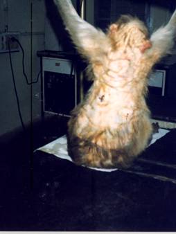
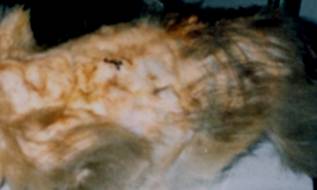
Figures 1 and 2 represent the animal with excessive hair loss from head to back regions with thickening and wrinkling of the skin, Scales and crust demarcate over the skin areas with erythamatous patches at varying degree. The animal's health looked apparently normal other than self-mutilation, itching and plucking its hair.
| Figure 1 and Figure 2 | |
|
|
| Figure-3 | 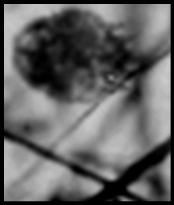 |
Sarcoptes Scabiei mites are ectoparasites in mammals and human being s but they have host preference although there is no remarkable morphological difference among mites of different hosts (Fainet.al.1968) .Zahler.et al reported that the genus sarcoptes consists of a single heterogenous species genotypically. Sarcoptes infection in humans (Sarcoptes scabiei var hominis) and sarcoptes scabiei was morphologically indistinguishable from each other (Mellanby, k, et al 1972,Fain A., et al 1968 and Kano, R., et al., 1999). Generally sarcoptes mites of an animal origin infected temporary and don’t lay eggs on humans (Alexander, J.O.D., et 1984). The mite burrows into the stratum corneum and feed on cells of stratum granulosum and stratum spinosum. Epidermal damage induces epithelial hyperplasia and the development of parakeratotic crust (YagerJ I.e. al 1993). Sarcoptes infection in animals is characterized by loss of hair, thickening and wrinkling of the skin and scab and crust formation. Severe infection may lead to changes of the skin and death of the animal and the hair become sparse with inflammation and irritation. Sarcoptes mites may develop crust, erythematous macules, and fissures over the skin with foul smelling wet crusted exudates above the head and in severe cases over the trunk and appendages.
After confirming the infection by both macroscopic and microscopic examination the animal was administered with Ivermectin at a dose level of 200 micrograms/kg body weight for 5 days (Graczyk et.al 2001) along with antihistamines and B-complex syrup as supportive therapy and gradual improvement of skin conditions were noticed after treatment.
Acknowledgement:
We are Thankful to Director NII for providing necessary facilities to carry out this case study.
References:
1..Alexander, J.O D .1984.Arthropods and Human Skin.Springer-V Verlag.
2.Graczyk, T.K, Mudakikwa A.B, CranField M.R, Eilenberger U.Hyperkerotic mange caused by Sarcoptes scabiei (Acariformes: Sarcoptidae) in Juvenile human –habituated mountain Gorillas (Gorilla gorilla berngei). Parasitology research. 2001,Dec:87(12):1024
3. Fain, A (1968.) Etude de la variabilite de Sarcoptes scabiei avec une revision des Sarcoptidae ActaZool.Pathol.Antverpiensia 47: 1-196
4.Kano, R.1999. Arthropods and Dermatology. Tokai University Press, Tokyo
5.Mellanby, k 1972. Scabies. . E W. Classey, Hampton
6.Scott WA (2003) Sarcoptic mange in Foxes. Vet Rec. 2003 Feb 8; 152(6): 183.
7.Smith E.K. (1988). How to detect common skin mites through skin scrapings. Vet. Med., 165-170.
8.Suzuki, Y., Sujimura, M.and Kaneko, K.1981. Res.Bull.Fac.Agri.Gifu Univ.45: 151-156
9.Yager J A, Scott DW: The skin and appendages in Pathology of domestic animals. Jubb KVF Kennedy PC, and Palmer N Eds. Academic press, San Diego, pp.681-682
10.Zahler, M., Essig, A., Gothe, R. and Rinder, H. (1999). Molecular analyses suggests monospecificity of the genus Sarcoptes (Acari: Sarcoptidae). Int. J. Parasitol. 29, 759-766.
Corresponding author ;
Dr.P.Nagarajan B.V.Sc ,Msc,
Veterinarian
Primate Research Center
National Institute Of Immunology
JNU Campus
New Delhi
India 110067
Phone # 91-26717129
Mail id: nagarajan @nii.res.in naga73@yahoo.com
| Click on these buttons to visit our journals | ||||||||
| Psychiatry On-Line | Dentistry On-Line | Vet On-Line | Chest
Medicine On-Line | GP On-Line | Pharmacy On-Line | Anaesthesia On-Line | Medicine On-Line | Family
Medical Practice On-Line |
All pages copyright ©Priory Lodge Education Ltd 1994-2004.
Copyright Priory Lodge Education Ltd 2004
First Published May 2004