CYTOLOGY
Fine Needle Aspirate Biopsies
by
Chris Belford BVSc MRCVS DVSc FACVSc
Fine needle aspirate biopsy FNAB is a relatively simple technique which
can be carried out to provide more information on a lesion.
3 Steps to Successful FNABs
1.Lesion Selection
Discrete, definable lesions provide the best results. Oedematous,
cystic, miniscule or extremely hard/fibrous (i.e. epulides) lesions may
not provide many ar any cells. Cells are of course what cytology relies
on!
2.Correct Sampling technique, preparation and speed
Speed of collection and sample preparation is very important to avoid
microclotting of the cells. Samples should be prepared immediately on
collection i.e. <0.5 seconds.
Here is the correct sampling technique and preparation:
- Use a 5 or 10ml syringe and a 21-25 gauge needle.
- Have a series of slides (3-5) waiting at arms length.
- Attach the needle to the syringe, pierce the lesion and withdraw the plunger to apply 3-5mls of vacuum.
- Keeping the vacuum constant and whilst remaining within the lesion at all times, redirect and "fan" the lesion
(as if you were attempting to infiltrate local anaesthetic) quickly, several times to obtain a representative
sample. This normally should only take a couple of seconds.
- Release the vacuum returning the plunger to zero and then remove the needle and syringe from the lesion.
Immediately detach the needle from the syringe, pull the plunger back to put 3-5mls of air into
the syringe and then reattach the needle.
- Depress the plunger quickly directing the air and sample via the needle onto the slides.
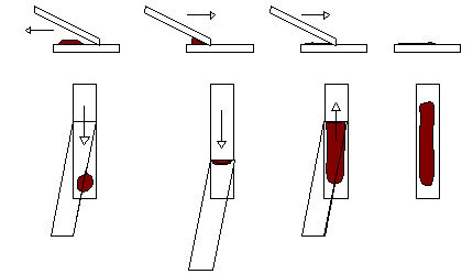
- Instantly make smears by the drawback and push away method as per blood
smears (avoid running the sample off the end of the slide- this is usually due to too much sample or low
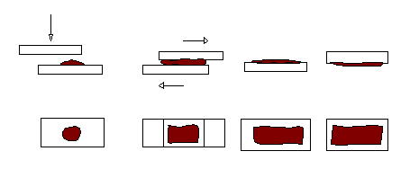
angle of the spreader slide) or by the pull apart method where one slide is placed on
top of the sample, and the slides pulled horizontally apart.
This method tends to rupture cells more frequently than the former method. However in mucinous samples
it may be required as the former method fails to produce adequate spreading.
- Wave smears actively to help rapid drying.
- Keep smears out of the fridge and away from formalin, even formalin fumes.
Figure 1
Drawback and Push Away Method

Figure 2
Pull Apart Method

Samples are best taken from a couple of sites within a lesion as lesions
are frequently not uniform. If the lesions are cystic, sample the edges
of the lesions, do not sample just the cystic fluid which may contain
few or no cells.
In the case of feline lymph nodes, vacuum should be kept to a minimum to
prevent cell rupture.
In the case of firm/fibrous lesions vacuum should be increased and
vigorous fanning performed to harvest adequate cells. Do not use a
larger gauge needle as it usually increases blood contamination and
needles larger than 21 gauge produce small core biopsies and not
individual cells.
3.Provide an adequate history and lesion distribution/descriptions on
sample submission for the cytologist.

Return to Vet On-Line Professional Pages
All pages copyright © Priory Lodge Education Ltd 1994-2000.


![]()