Walter Tarello, DVM
C.P. 1644
06129 PERUGIA (ITALY)
This work has been carried out at the 'Heron Veterinary Clinic' of Castiglione
del Lago (Perugia province) and in a private veterinary practice of Fermo (Ascoli
Piceno province), Italy.
Current affiliation: Ambulatorio Veterinario Spina, Piazza Vittoria, Spina (Perugia).
SUMMARY
Background: Lack of notions on distribution and pathogenicity of D. repens
among cats living in Central Italy suggested this study. Main results: Pruritic
dermatitis associated with D. repens microfilariaemia was diagnosed in 19 cats
from the Tuscany, Umbria and Marche regions of Italy. Dermatological lesions
included alopecia, erythema, papulae, crusting and lichenification. All cats
had lesional pruritus and 90% had concurrent haemobartonellosis. Eradication
of haemobartonellosis followed by specific adulticide and microfilaricide treatment
led to disappearance of microfilaraemia and complete recovery from the dermatological
syndrome. Implications: The main conclusion is that cats are reservoir for D.
repens in Central Italy and constitute a possible source of accidental infestation
for humans. Furthermore, the cutaneous lesions observed in these cases seem
to attribute a pathogenic role to D. repens in affected cats.
INTRODUCTION
Dirofilaria repens is a zoonotic filarial nematode parasite of dogs, cats and
wild carnivores transmitted by mosquitoes (Pampiglione et al., 1995). Adult
worms reside in the subcutaneous tissues of infested animals and release microfilariae
that circulate in the blood (Tarello, 1999). Dogs and cats are final hosts of
the nematode, which also accidentally affects people, causing sub-cutaneous,
conjunctival and pulmonary nodules often confused with neoplastic tumors (Pampiglione
and Rivasi, 2000). Italy has the highest concentration in the world of recorded
cases of human dirofilariasis due to D. repens (Pampiglione et al., 2001). Nonetheless,
to date, no data are available on the presence of microfilaraemia and possible
pathogenicity due to Dirofilaria repens in cats living in Central Italy.
The geographical distribution of human cases follows the distribution of microfilaraemia
observed in dogs and cats (Pampiglione et al., 1995; Tarello 2002a-b).
It was thus worthy to report this retrospective analysis of 19 feline cases
of subcutaneous dirofilariasis observed in an area of Central Italy, from wich
4 human cases have already been reported (Pampiglione et al., 1995; Pampiglione
and Rivasi, 2000) and suitable vectors are present (Romi et al., 1997; Grelloni
and Biasini, 2002), although no animal reservoirs has ever been noticed.
METHODS
Fourteen cats from the Trasimeno lake district (Perugia province, Umbria), two
cats from the neughboring Chiusi (Siena province, Tuscany) and three cats from
Fermo (Ascoli Piceno province, Marche) were presented at the author's practices
during 1997 (Trasimeno and Chiusi) and 1999 (Fermo) for pruritic dermatitis.
Laboratory tests for Dirofilaria repens and Dirofilaria immitis microfilariae
were carried out using a modified Knott's test on 1 milliliter whole blood sample
collected in EDTA tube. Six microscopical preparations were made from each sample
and examined at light microscopy (x4 and x10, Leitz Biomed).
Serological testing for circulating antibodies to Dirofilaria immitis was carried
out with a commercial kit (HeskaTM Feline Heartworm, Heska Corporation). A Wright-stained
fresh blood smear was also done for each cat to check for other feline haemoparasites.
Informations on the history, duration of the disease, previous treatments, dermatological
and general clinical signs and the results of blood tests were collected in
each case.
Diagnosis of subcutaneous dirofilariasis was based on the following established
criteria: a) pruritus lasting more than 1 wee; b) presence of cutaneous lesions;
c) microfilariasis caused only by Dirofilaria repens; d) negative serology for
antibodies to Dirofilaria immitis.
No cat had received preventive medication for heartworm disease and no cat was
presented with flea infestation at the time of consultation.
Therapy was based on the preventive eradication of the concurrent Haemobartonella
felis infection with doxycycline at anti-rickettsial dosage (Ronaxan 20, Merial,
10 mg/Kg., os., for 20 days) followed by a treatment with the arsenic adulticide
melarsomine (Immiticide, Merial, 2.5 mg/Kg, im., twice, q. 24 hours) and the
microfilaricide drug ivermectin (Ivomec, Merial, 50 m/Kg, sc., once) given ten
days after the treatment for the macrofilariae. Follow-up included a clinical
re-examination and a Knott test one month after the end of the treatment.
RESULTS
Diagnosis of subcutaneous dirofilariasis due to Dirofilaria repens was made
on 19 cats from Central Italy, the age of which ranged between 8 months and
12 years. Thirteen were males and six females. At the time of inspection the
condition lasted 2 weeks-3 years. Previous unsuccessful non-specific antipruritic
treatments, flea control and restricted diet were recorded in 9 cats (47.3%).
All cats had pruritus, manifested by localised scratching, licking and biting.
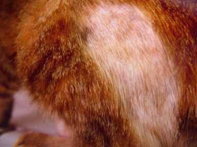
The gross appearance of skin lesions was as follows: focal or multifocal alopecia
(n = 17; 90%) (Fig. 1), erythema (n = 15; 79%), papulae (n = 11; 57.9%), crusting
(n = 5; 26.3%) and lichenification (n = 2; 10.5%). Symptoms and signs other
than dermatological were as follow: anorexia (n = 7; 36.8%), pale mucous membranes
(n = 6; 31.6%), lethargy (n = 4; 21%), lymph-adenomegaly (n = 4; 21%), hyperestesia
(n = 4; 21%), fever (n = 3; 15.8%) and conjunctivitis (n = 2; 10.5%).
Microscopic examination of six slides from every Knott concentration test was
carried out and Dirofilaria (Nochtiella) repens microfilariae were found in
the blood of all cats (Fig 2), their count ranging from 5 to 17 per sample.
Dirofilaria immitis microfilariae were not found and the serologic antibodies
test HeskaTM FH resulted negative. Concurrent Haemobartonella felis infection
was diagnosed in 17 cases (90%).
All patients recovered after specific treatment against both the haemobartonellosis
and the dirofilariasis. Clinical re-examination and a Knott test, carried out
1 month after the completion of treatment, showed the resolution of the cutaneous
lesions and the absence of Dirofilaria repens microfilariae in the blood.
DISCUSSION
Nineteen cats from Tuscany, Umbria and Marche regions of Italy affected by pruritic
dermatitis (Fig. 1) were found to carry Dirofilaria repens microfilariae in
the blood (Fig. 2), confirming that domestic felines are a valuable reservoir
for infestation to humans in Central Italy. Suitable vectos exist (Romi et al.,
1997), including Aedes albopictus (Grelloni and Biasini, 2002), and four human
cases have been reported in the area, three from Siena (Pampiglione et al.,
1995, p.173-174) and one from Perugia province (Pampiglione and Rivasi, 2000,
p. 237) indicating that the infestation was acquired locally. In fact, history
of travel to endemic areas was absent from the medical descriptions of these
cases.
The importance of such findings resides in the fact that no dog, cat or wild
carnivore from these two provinces (Siena and Perugia) has ever
been found to carry microfilariae of D. repens, although suitable animal reservoirs
shoul exist in sites where human dirofilariasis is repeatedly and not occasionally
acquired or imported (Pampiglione and Rivasi, 2000).
The observations here reported may permit a risk assessment in front of a possible
transmission to humans, since cats are more linked to the territory than dogs
and consequently can provide better epidemiological indications. Only two Dirofilaria
spp. (immitis and repens) affect cats (Tarello, 2000a; Kramer and Genchi, 2002)
and differentiation is mainly based upon the morphology of the microfilariae
(Tarello, 2001). Exclusion of D. immitis, the causative agent of heartworm disease,
through a negative antibodies test was an important confirmation that microfilariae
observed were only due to D. repens.
In fact, circulating antibodies to D. immitis are present not only during infection
with adult worms, but also during infection with immature and larval stages
(Kramer and Genchi, 2002).
Adult nematodes of D. repens are usually localized in the subcutaneous tissues
(Pampiglione et al., 1995; Chauve, 1997) and were not found, nor were they searched
for, during this study. Nonetheless, the detection of microfilariae in the blood
seems to be of significant diagnostic importance for the presence of adults
in Dirofilaria infestations (Anon, 1998).
Dirofilaria repens has never been reported in animals from Umbria region although
it is considered endemic in dogs living on seacosts of Tuscany (Tarello, 1999)
and Marche (Tarello, 2002a).
Nonetheless, in all these regions of Central Italy no feline case has ever been
described. On this account, it is particularly true the observation that absence
of evidence is not equivalent to evidence of absence. No specific reason, in
fact, might exclude the parasitosis in cats in areas where human cases are reported,
since both dog and cat are recognized reservoirs and definitive hosts of this
zoonotic nematode in endemic areas of the world (Mak et al., 1980; Pampiglione
et al., 1995; Olteanu, 1996; Dissanaike et al., 1997; Tarello, 2000a-b and 2002a-b).
Cats of this study live in rural areas and were allowed access to outdoor environments.
This fact enhances the risk of mosquitoes bite and consequently the risk of
acquire Dirofilaria spp. infestations (Kramer and Genchi, 2002).
Epidemiology of feline subcutaneous dirofilariasis has received little attention
in the past, if compared with studies carried on the canine population (Rossi
et al., 1996).
Microfilaraemia (Fig. 2) has been observed in cats from Malaysia (Mak et al.,
1980), Sri Lanka (Dissanaike et al., 1997), Romania (Olteanu, 1996) and Italy
(Pampiglione et al., 1995), all nations where human cases are recorded (Pampiglione
and Rivasi, 2000).
Acute liver failure in a cat has been recently reported from South Africa (Schwan
et al., 2000) and skin lesions associated with D. repens microfilariae in the
blood have been observed in 11 cats living in highly endemic area of Northern
Italy (Tarello, 2000a-b).
The cases reported in this study, showing itching dermatitis (Fig. 1) responsive
to specific medicaments, seem to confirm a pathogenic role
for D. repens in animals, previously disputed (Chauve, 1997).
Erythema and pruritus are described also in human cases, although microfilaraemia
is absent and usually only one adult parasite is recovered (Pampiglione and
Rivasi, 2000; Pampiglione et al., 2001).
On the other hand, haemobartonellosis, the concurrent condition observed in
90% of cats in this study, is not clinically associated with pruritus and cutaneous
lesions (Baneth et al., 2002). Treatment of subcutaneous dirofilariasis has
been rarely reported in cats (Tarello, 2000a-b) although it is suggested for
animals suffering from clinical signs of this disease (Baneth et al., 2002).
Therapy of the condition is important in order to decrease the risk of infestation
to humans in the vicinity of the affected animal when suitable mosquito vectors
are present (Baneth et al., 2002).
Dirofilariasis due to D. repens is an emerging zoonosis in Italy (Pampiglione
et al., 2001) and it is acknowledged that worldwide distribution of human cases
follows the distribution of microfilaraemia observed in dogs and cats (Pampiglione
et al., 1995).
The findings here reported seem to indicate that a feline reservoir for the
nematode exist in this area of Central Italy. Lack of previous evidence may
be due to the fact that during the last years the ranges of certain vector-borne
diseases such as dirofilariasis are extending due to ecological and climatic
changes (Irwin, 2002). Involvement of Aedes albopictus may also account for
such expansion (Grelloni and Biasini, 2002).
Recommendation has been recently made that each and every case of human dirofilariasis
observed in Italy should be recorded, in order to determine the true extent
of the condition (Pampiglione et al., 2001).
In companion animal practice, veterinarians have the responsibility of providing
accurate information about the zoonotic transmission of parasite infections
from pets (Irwin, 2002). In this view, canine and feline cases of subcutaneous
dirofilariasis should be also reported as far as possible, in order to assess
the presence, prevalence and pathogenicity of Dirofilaria repens in the animal
reservoirs and final hosts.
REFERENCES
Anon. (1998) Heartworm disease. J. Small Anim. Pract. 39, 407-408.
Baneth G, Volansky Z, Anug Y, Favia G, Bain O, Goldstein RE, Harrus S. (2002) Dirofilaria repens infection in a dog: diagnosis and treatment with melarsomine and doramectin. Vet. Parasitol. 105, 173-178.
Chauve CM. (1997) Importance in France of the infestation by Dirofilaria (Nochtiella) repens in dogs. Parassitologia. 39, 393-395.
Dissanaike AS, Abeyewickreme W, Wijesundera M de S, Weerasoorija MV, Ismail MM. (1997) Human dirofilariasis caused by Dirofilaria (Nochtiella) repens in Sri Lanka. Parassitologia. 39, 375-382.
Grelloni V, Biasini G. (2002) Detection of Aedes albopictus in the province of Perugia (Umbria). Experimental Zooprophylactic Institute of Umbria and Marche, Report n. 49799 of 19 August 2002 (see picture n. 4 enclosed).
Harrus S, Klement E, Aroch I, Stein T, Bark H, Lavy E, Mazaki-Tovi M, Baneth G. (2002) Retrospective study of 46 cases of feline haemobartonellosis in Israel and their relationship with FeLV and FIV infections. Vet. Rec. 151, 82-85.
Irwin PJ. (2002) Companion animal parasitology: a clinical perspective. Int. J. Parasitol. 32, 581-593.
Kramer L, Genchi C. (2002) Feline heartworm infection: serological survey of asymptomatic cats living in northern Italy. Vet. Parasitol. 104, 43-50.
Mak JW, Yen PK, Lim KC, Ramiah N. (1980) Zoonotic implications of cats and dogs in filarial transmission in Peninsula Malaysia. Trop. Geogr. Med. 32, 259-264.
Olteanu G. (1996) Dirofilariosis in man and animals in Romania. Parassitologia. 38, 360.
Pampiglione S, Canestri-Trotti G, Rivasi F. (1995) Human dirofilariasis due to Dirofilaria repens. Review of the world literature. Parassitologia. 37, 149-194.
Pampiglione S, Rivasi F. (2000) Human dirofilariasis due to Dirofilaria (Nochtiella) repens: an update of world literature from 1996 to 2000. Parassitologia. 42, 231-254.
Pampiglione S, Rivasi F, Angeli G. (2001) Dirofilariasis due to Dirofilaria repens in Italy, an emergent zoonosis: report of 60 new cases. Histopathology. 38, 344-354.
Romi R, Pierdominici G,
Severini C, Tambutto A, Cocchi M, Menichetti D, Pili E, Marchi A. (1997) Status
of malaria vectors in Italy. J. Med. Entomol. 34, 267-271.
Rossi L, Pollono F, Meneguz PG, Gribaudo L, Balbo T. (1996) An epidemiological
study of canine filarioses in North-west Italy: what has changed in 25 years?
Vet. Res. Comm. 20, 308-459.
Schwan EV, Miller DB, De Kock D, Van Heerden A. (2000) Dirofilaria repens in a cat with acute liver failure. J. South Afr. Vet. Ass. 71, 197-200.
Tarello W. (1999) La dirofilariose sous-cutanee a Dirofilaria (Nochtiella) repens chez le chien. Revue bibliographique et cas clinique. Revue Med. Vet.150, 691-702.
Tarello W. (2000a) La dirofilariose sous-cutanee a Dirofilaria (Nochtiella) repens chez le chat: symptoms, diagnostic et traitement sur 10 cas. Revue Med. Vet. 151, 813-819.
Tarello W. (2000b) Un cas de dirofilariose sous-cutanee chronique a Dirofilaria (Nochtiella) repens chez un chat. Revue Med. Vet. 151, 969-971.
Tarello W. (2001) Importance in the dog of concentration tests for the diagnosis of heartworm disease in non endemic areas. Vet On-Line, 02 Jan 01: http://www.priory.com/vet/cardioworm.htm.
Tarello W. (2002a) Dermatitis associated with Dirofilaria (Nochtiella) repens microfilariae in dogs from Central Italy. Acta Vet. Hung. 50, 63-78.
Tarello W. (2002b) Cutaneous lesions in dogs with Dirofilaria (Nochtiella) repens infestation and concurrent tick-borne transmitted diseases. Vet. Dermatol. 13, 267-274.
FIGURE LEGEND
Fig 1) Focal alopecia in a cat with D. repens infestation.
Fig. 2) Dirofilaria repens microfilaria in the blood of a cat.
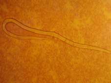
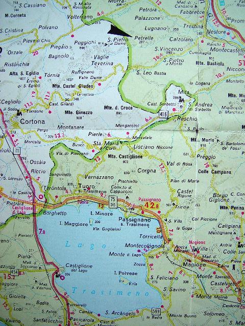
Fig. 3) Map showing the working area (Trasimeno Lake District) and the site
(arrow = Petrelle) in which the first human case of dirofilariasis has been
recently discovered.
Submitted November 2002
First Published November 2002
| Click on these buttons to visit our journals | ||||||||
| Psychiatry On-Line | Dentistry On-Line | Vet On-Line | Chest
Medicine On-Line | GP On-Line | Pharmacy On-Line | Anaesthesia On-Line | Medicine On-Line | Family
Medical Practice On-Line |
All pages copyright ©Priory Lodge Education Ltd 1994-2004.