Early in 1995, the kennel population of more than 100 dogs at the Guide Dogs for the Blind Association's Midlands Regional Centre in Leamington Spa was hit by an unusually severe outbreak of diarrhoea. The cause turned out to be an infection of the intestine by a commonly-occuring, single celled organism - or protozan known as Giardia. A combined treatment and disinfection strategy was then introduced that brought the infection under control.
Maggie Fisher, a veterinary surgeon with a special interest in parasitology, was called in to help deal with
the Giardia outbreak, and in the following paper she describes the infection and how it can be treated and controlled
The division of Giardia into groups according to species is still somewhat confused; the organisms that infect mammals look very similar but it remains unclear to what extent they form one or a number of species. It is for this reason that, while Giardia infection in some mammals, including dogs, is suspected of being infectious to man (ie: a zoonosis), it has not been conclusively shown that the species in, for example, dogs and man is the same.
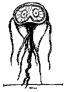
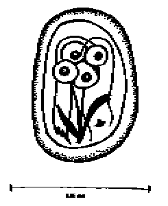
The Giardia trophozoite (Figure 1) - which is the active stage of the organism - inhabits the small intestine of the dog. It attaches to the cells of the intestine with its adhesive disc and rapidly divides to produce a whole population of trophozoites. As they detach they may be swept down the intestine. If intestinal flow is fast then they may appear in the faeces. However, if they have time, they will develop into the inactive, more durable, cyst form of the organism and these will be passed in the faeces. The cyst (Figure 2) is more able to survive in the environment than the trophozoite, which is very fragile.
The cysts are infective as soon as they are passed, unlike other parasites where a lag period is necessary before the organism is infective. The most common route of infection is faeco-oral. For example, dogs may accidentaly eat cysts as they lick around theenvironment or lick other dogs' coats (particularly if the other dog has diarrhoea). Another major source of infection in human cases is drinking contaminated water. Once eaten, the cyst breaks open in the animals' intestine and releases two new trophozoites to initiate infection. If a dog is left in a dirty environment it may act as its own source of further infectionas it eats cysts passed in its own faeces.
The elisa technique requires a kit and some method of reading a colour change or production of flourescence. Studies examining the reliability of some immunoflourescent kits have found them to be over 90% accurate, with relatively few false negatives or false postives. However, the tests are costly and probably only wothwhile where there are alarge number of samples to be processed and a technician who is familiar with carrying out elisas.
Treatments for Giardias in dogs | |||
|---|---|---|---|
| Drug Name | Trade Name | Dose Rate | Duration of Treatment |
| Metronidazole | Flagyl | 25-30 mg/kg bid** | 7 days |
| Furazolidone | Neftin | 4 mg/kg bid* | 10 days |
| Tinadazole | - | 44 mg/kg once daily | 7 days |
| Fenbendazole | Panacur *** | 50 mg/kg once daily | 3 days |
| Albendazole | Valbazen | 25 mg/kg bid | 2 days |
| bid | Twice daily |
| * | Maximum daily dose 200 mg |
| ** | Contra-indicated in pregnancy |
| ** | Licenced for the treatment of worm infections in dogs |
Option 1 is only practical where a few dogs in a discrete area have been identified as being infected and where complet isolation is feasible, either within their own block or in a specific isolation block. Such isolation includes segregation of exercise areas and thes animals should be fed and cleaned after all others on the premises, preferably using separate cleaning and feeding equipment and separate staff if possible. Treatment of all dogs should commence on the same day when option 2 is adopted.
Thorough cleaning of all kennel area where infected dogs have access is essential. Once organic debris has been removed, thorough disinfection will help to further reduce the level of environmental contamination and reduce the risk of dogs becoming re-infected after the completion of treatment. Disinfectants containing quaternary ammonium compounds have been found to kill Giardia cysts at the manufacturers' recommended dilutions (dilutions of one disinfectant upto 1:704 were found to be effective at both low and high environmental temperatures). Efficacy of killing is increased by prolonged contact time, therfore disinfectant solution should be left for 20 minutes to half an hour before being rinsed off kennel or run surfaces. Since disinfection of grass runs is impossible, such area should be regarded as contaminated for atleast a month after infected dogs last had access.
Introduction of new dogs into the infected area should be avoided until the period of treatment and faecal samle checking has been completed. It should not be overlooked that some of thoe infected dogs may continue to excrete low numbers of cysts even after all treatments and examinations have been completed. It is therefore important that rigorous disinfection is maintained and a careful check is kept on the condition of all treated and introduced animals.
Dogs should be prevented from access to foul water that may contain large numbers of cysts (eg: river-flooded paddocks).Small numbers of cysts may occasionally be present in the potable water supply but the risk of this being a major source of infection is small.
![]()
Return to Vet On-Line Professionals
All pages copyright ©Priory Lodge Education Ltd 1994-2000.