Sheila C. Rahal, Lígia S. L .S. da Mota, Maria J. Mamprim, Renata B.
Ciani
Department of Veterinary Surgery and Anesthesiology (Rahal, Ciani) and Department of Animal Reproduction and Radiology (Mamprim), Faculty of Veterinary Medicine and Animal Science - UNESP Botucatu, Caixa Postal 560 - Rubião Júnior, s/n, CEP: 18618-000 - Botucatu (SP) - Brazil; Department of Genetic (Mota), Bio science Institute - UNESP Botucatu.
Address correspondence to Dr. Sheila C. Rahal
Case Report
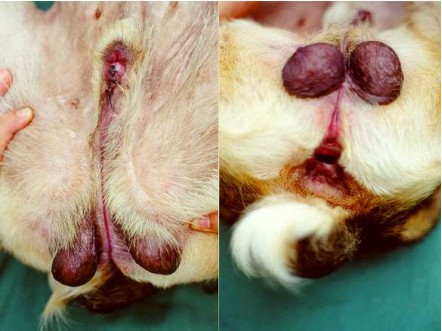
An eight-month-old, crossbreed dog, weighing 9 kg, was admitted to the Veterinary
Hospital with congenital abnormalities of the urogenital tract. It was the only
animal of the litter with deformity. On physical examination, underdevelopment
of the penis, fusion failure of the urethra, prepuce and scrotum (Figure 1a),
and an external urethral orifice located in the perineum about 1.2 cm below
the anus (Figure 1b), were observed. Testicles could be palpated in the scrotal
area. A groove covered by mucosa extended cranially from the cleft scrotum to
the glans penis area. The dog did not display dysuria or urinary incontinence,
but he squatted to urinate.
Alteration of the urinary bladder shape with dilatation of its neck and presence of ventral diverticulum were observed on ultrasonographic examination (Figure 2a) and retrograde cystourethrography (Figure 2b).
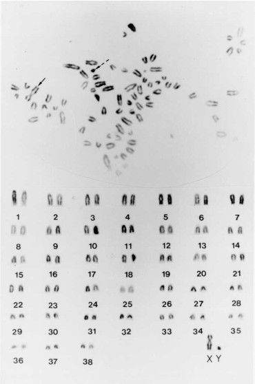
Chromosome analysis was performed. Peripheral blood samples and tissue were cultured as described (1, 2). Sixty cells were cytogenetically analyzed and the chromosomes were identified and classified according to Mellink and Bosma (1989) (3). Normal karyotype (78, XY) was found in all mitotic metaphases cells analyzed by Giemsa staining (Figure 3). The diagnosis was perineal hypospadias.
Discussion
Hypospadias is a congenital anomaly of the external genitalia, in which there
is fusion failure of the urogenital folds and incomplete development of the
penile urethra (4, 5, 6). The condition is rare and the etiology is unclear
(7). The types are glandular, penile, scrotal, perineal, and anal according
to the place of the urethral opening (4, 6). It can be classified as mild, moderate
or severe (8). In severe cases, lesions such as underdevelopment or absence
of the penis, failure of fusion of the scrotum, and failure of the urethra to
close in the perineal area may be seen, similar to the case reported here (4,
9).
Other abnormalities associated with hypospadias such as retained testicles, kidney agenesis, bone or anorectal defects, umbilical hernia, hydrocephalus, and urinary incontinence (7, 10, 11) were not detected in this case. Urinary incontinence also has been observed in three reported cases (9). Chromosome analysis can be used to differentiate hypospadias from true hermaphroditism (9). This confirmed that the animal in this case report was a male.
No corrective surgery is indicated with minimal defects, moderate urethral defects can be treated with a two-layer closure, and major defects with excision of the external genitalia and urethrostomy (4, 9). Surgery was declined in this case. In spite of the deformity, the animal did not show relateed health problems and did not show urine scalding of the skin. Animals with this defect may show ascending urinary tract infections, but none were evident in this case (5).
Figure 1. Perineal hypospadias. Observe underdevelopment of the penis, fusion failure of the urethra, prepuce and scrotum (a), and external urethral orifice located in the perineum (b).
Figure 2. Ultrasonography (a) and
retrograde cystography (b) showing urinary bladder with ectopia and alteration
of the shape.
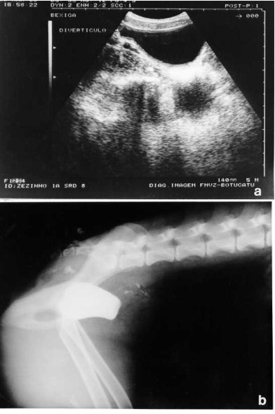
Figure 3. Karyotype and mitotic metaphase chromosomes obtained from tissue culture after conventional Giemsa staining. The continuous arrow indicates the X chromosome and the dashed arrow Y chromosome.
1. Moorhea PS, Nowell PC, Nellman, WJ, Battips, DM, Hungerford,
DA. Chromosome preparations of leukocytes cultured from human peripheral blood.
Exp Cell Res 1960; 20: 613-616.
2. Freshney RI. Culture of animal cells. A manual of basic technique. 3rd ed.
New York: Wiley-Liss. 486p.
3. Mellink CNM, Bosma AA. The karyotype of the domestic dog (Canis familiaris
L.). In: Halnan CRE. Cytogenetics of animals. Wallinford: CAB International,
1989: 151-157.
4. Hobson HP. Penis and prepuce. In: Bojrab MJ, Ellison GW, Slocum B. Current
Techniques in Small Animal Surgery. 4th ed. Philadelphia: Lea & Febiger,
1998: 527-537.
5. Smith CW. Surgical diseases of the urethra. In: Slatter DH, ed. Textbook
of Small Animal Surgery. 2nd ed. Philadelphia: WB Saunders, 1993:1462-1463.
6. Fossum TW, ed. Small Animal Surgery. St. Louis: Mosby, 1997: 565-566.
7. Hayes HM, Wilson GP. Hospital incidence of hypospadias in dogs in North America.
Vet Rec 1986,118:605-607.
8. Hoskins JD. Congenital defects of the dog. In: Ettinger SJ, Feldman EC. Textbook
of Veterinary Internal Medicine. 4th ed. Philadelphia: WB Saunders, 1995: 2126.
9. Ader PL; Hobson HP. Hypospadia: a review of the veterinary literature and
a report of three cases in the dog. J Am Anim Hosp Assoc 1978; 14:721-727.
10. Hobson HP. Surgical pathophysiology of the penis. In: Bojrab MJ, Smeak DD,
Bloomberg, MS. Disease mechanisms in small animal surgery. 2nd ed. Philadelphia:
Lea & Febiger, 1993: 554-555.
11. McFarland LZ, DENIZ E. Unilateral renal agenesis with ipsilateral cryptorchidism
and perineal hypospadias in a dog. J Am Vet Med. Assoc 1961; 139:1099-1100.