This article aims to describe atopic dermatitis and its treatment in the 1990's, and draws attention to some of the important questions yet to be answered.
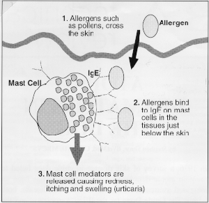 Antibodies are of several
types, IgG for instance being involved in protection against viral diseases after vaccination whereas IgE,
involved in atopic dermatitis, is particularly concerned with protection against parasites. IgE antibodies
coat specialised cells (mast cells) in the skin where they sit waiting for contact with the parasite proteins
to which the animal is sensitised. If the substance is encountered, perhaps as a result of a burrowing mite,
the mast cell releases chemicals (mast cell mediators) which try to destroy the invader. In allergic animals
this whole system is oversensitive and the release of mast cell mediators in the skin occurs inappropriately
to apparently innocuous substances such as pollens, moulds and house dust mites (figure 1).
Antibodies are of several
types, IgG for instance being involved in protection against viral diseases after vaccination whereas IgE,
involved in atopic dermatitis, is particularly concerned with protection against parasites. IgE antibodies
coat specialised cells (mast cells) in the skin where they sit waiting for contact with the parasite proteins
to which the animal is sensitised. If the substance is encountered, perhaps as a result of a burrowing mite,
the mast cell releases chemicals (mast cell mediators) which try to destroy the invader. In allergic animals
this whole system is oversensitive and the release of mast cell mediators in the skin occurs inappropriately
to apparently innocuous substances such as pollens, moulds and house dust mites (figure 1).
For allergy to be apparent, dogs need to be first "allergic" and then be exposed to substances (allergens) to which they can develop the abnormal immune response. In the UK the main source of allergens is the house dust mite. These tiny creatures live in all of our houses, in carpets, beds and other soft furnishings and feed on skin scales that are constantly falling from people and animals. They litter our environments with fćcal pellets of half-digested food and digestive enzymes and it is these minute faecal particles that contain the most important allergens. Dogs can also become allergic to pollens and moulds although this is much less common, presumably because of less exposure.
Other factors known to be important in atopy in man are certain infectious diseases in the early part of life which modify the response to allergens. In particular it has been shown that children who have more respiratory infections early in life, before any allergy is apparent, have a lower chance of showing signs of allergy. The effect of such infections is not known in the dog.
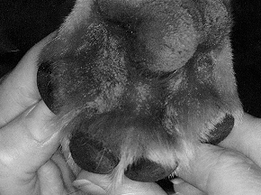 Luckily there are some tell-tale
signs that help us to identify the pruritic dog. Saliva staining is a commonly-seen feature in these animals.
A red-brown staining of light coloured hair is often seen in allergic dogs in the groin, armpits(axillae) and
between the toes(interdigital spaces)and can be seen in figure 2. In addition, with long term
problems, the skin itself will also change colour. Instead of being pink, a black mottling
(hyperpigmentation) will slowly develop, especially if the skin has looked red and angry at the site. This is
most commonly seen on the abdomen.
Luckily there are some tell-tale
signs that help us to identify the pruritic dog. Saliva staining is a commonly-seen feature in these animals.
A red-brown staining of light coloured hair is often seen in allergic dogs in the groin, armpits(axillae) and
between the toes(interdigital spaces)and can be seen in figure 2. In addition, with long term
problems, the skin itself will also change colour. Instead of being pink, a black mottling
(hyperpigmentation) will slowly develop, especially if the skin has looked red and angry at the site. This is
most commonly seen on the abdomen.
So what are these other diseases? Flea infestation and the allergy are the most important causes of itchiness in dogs in the UK. Practically all dogs will have fleas at some time during their lives. The rump and hind end are most often affected. Nibbling and itching gives a rough feel to the coat and, if severe, pyotraumatic dermatitis ( wet eczema) or alopecia will result. Very importantly, dogs with atopic dermatitis are often allergic to fleas as well, so it is pointless making a diagnosis of atopy without taking rigorous flea-control measures. Similarly, other parasitic infestations such as lice or sarcoptic mange may mimic atopy and these should be carefully ruled out.
Food sensitivity ( often called food allergy) is an uncommon cause of allergic skin disease, which
accounts for a small percentage of the cases seen by dermatologists. Although a rare condition, all
allergic dogs should undergo food trials before being committed to long-term drug therapy. Food
sensitivity may coexist with atopy or flea allergy and so partial responses may be seen to food changes.
Bacterial infections are a common cause of pruritus in the dog and these can be as a result of atopic
dermatitis or any other skin condition that damages the integrity of the skin. Non-allergic causes of
bacterial infection include hormonal problems such as hypothyroidism and parasitic problems such as
demodex infestation. These are normally non-itchy conditions, but as soon as there is bacterial
involvement this changes and it can be difficult to make the correct diagnosis.
 Skin testing is performed to identify the allergens involved in allergic disease. Under profound sedation
an area of hair on the chest is shaved and small injections of substances known to be possible allergens
made. After 15-20 minutes the reactions are recorded. Figure 3 shows positive reaction to house
dust mite allergens in an allergic Labrador Retriever.
Skin testing is performed to identify the allergens involved in allergic disease. Under profound sedation
an area of hair on the chest is shaved and small injections of substances known to be possible allergens
made. After 15-20 minutes the reactions are recorded. Figure 3 shows positive reaction to house
dust mite allergens in an allergic Labrador Retriever.
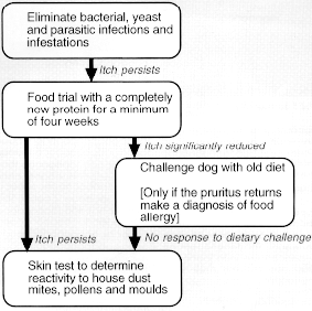
The diagnostic approach to the pruritic dog is summarised in figure 4.
 In treating atopic dermatitis it is imperative to consider the situation as a whole. Bacterial infections will
make the animal far more itchy and may even contribute to worsening the allergy through damaging
the skins' protective mechanisms. So any bacterial infections seen as a rash or pustular spots
(Figure 5),
need to be treated promptly, using a combination of shampoos and antibiotics for a minimum of three
weeks, and often longer. Corticosteroid medication is best withdrawn throughout the period of treatment
as steroids can interfere with the dogs ability to fight infection.
In treating atopic dermatitis it is imperative to consider the situation as a whole. Bacterial infections will
make the animal far more itchy and may even contribute to worsening the allergy through damaging
the skins' protective mechanisms. So any bacterial infections seen as a rash or pustular spots
(Figure 5),
need to be treated promptly, using a combination of shampoos and antibiotics for a minimum of three
weeks, and often longer. Corticosteroid medication is best withdrawn throughout the period of treatment
as steroids can interfere with the dogs ability to fight infection.
Yeast infection ( caused by the yeast Malassezia pachydermatis) is another complication. Spots are not seen in this disease, but instead the organism causes redness, geasiness and a mousy odour. Dogs can be quite depressed when infected and can be extremely itchy. Treatment is usually with baths containing enilconazole, or miconazole in combination with chlorhexidine. Tablet therapy is also available, but as a surface infection Malassezia is best treated using baths.
Similarly, fleas and other ectoparasites will make an atopic dog far more itchy. All allergic animals should have regular and efficient flea therapy using veterinary preparations to treat both the dog and the environment. With bacterial, yeast and parasitic problems under control most dogs will be very much more comfortable and some may only need minimal therapy using the least potent of the drugs available.
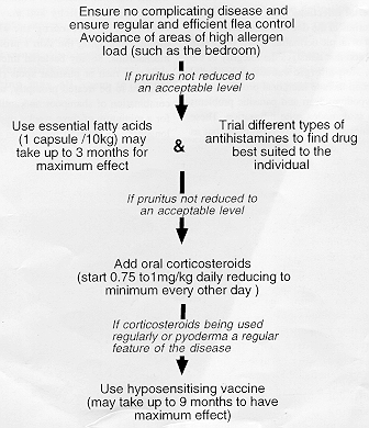
Essential fatty acids are now widely used for skin conditions. They are known to have few side effects and will help about 25% of allergic dogs significantly. Antihistamines potentiate the action of essential fatty acids (synergy) and so combination therapy would appear to be valuable. Several veterinary products are licensed for use.
Antihistamines were widely dismissed as unhelpful in atopic disease until recently when new studies both in the UK and USA have shown considerable benefits from their use. No veterinary products are available and the human drugs , chlorpheniramine, hydroxyine, and clemastine have all shown to be useful.
Steroids are widely thought to cause side effects which outweigh their potential for good. Despite this popular view, steroids are the drug of choice in severe cases of atopic dermatitis and, used appropriately, when complicating diseases are under control, side effects are generally minimal.
Hyposensitising Vaccines ( also known as desensitising vaccines) are prepared from the allergens identified as important at skin test. By administering these allergens subcutaneously over a long period the immune response to them is modified and pruritis is reduced. They are seen to be beneficial in about 60% of dogs, and take up to nine months to have effect.
Allergen avoidance is useful when house dust mites are known to be the problem. Exposure to bedrooms should be avoided by house dust mite allergenic patients to minimise exposure to the allergen. When pollens and moulds are involved avoidance is practically impossible as these allergens travel for miles on the wind, although obviously very large sources of pollens, for instance hay meadows for grass sensitive individuals should be avoided.
The author Stephen Shaw, is Dermatology Research Fellow at the Animal Health Trust in Newmarket, Suffolk, England, which involves him in clinical and research work with The Guide Dogs for the Blind Association (GDBA). The GDBA manages a breeding stock of about 250 dogs and raises 900 puppies every year. It also supports programmes aimed at improving the health and welfare of more than 6000 dogs for which it is responsible, and the quality of service for over 4000 guide dog owners throughout the UK.
![]()
Return to Vet On-Line Professionals
All pages copyright ©Priory Lodge Education Ltd 1994-2000.