Ascariasis - A case report
Kumar
& Banerjee
Case History:
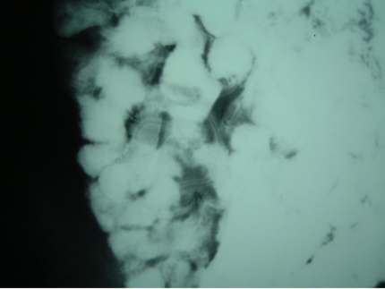
A 32 year old gentleman was referred to the radiology department for a Barium Meal Follow Through examination. The film shown here is one from the series. The patient presented with pain abdomen. An ultrasound and gastroscopy examination was negative. Inspite of everything the pain was still persisting. The patient is otherwise healthy.

Questions:
1)
What is the abnormality on this film?
2) Can this cause abdominal pain?
3)
What causes this?
4) How to confirm this?
5) What is the treatment for this
condition?
6) Are there any complications?
Answers:
1)
There is tram track appearance and filling defect noted which is marked and is
suggestive of Ascariasis.
2) Ascariasis infestation can cause pain and even
obstruction.
3) Ascariasis is the most common infestation that is caused by
Ascaris Lumbricoides.
4) To confirm this condition, stool examination is important.
In this patient he stool examination confirmed the presence of mature worm in
the faeces. The blood examination might show Eosinophelia and in some cases the
chest x-ray may exhibit some alveolar shadowing, which spontaneously clears.
5)
The treatment includes antihelminthic drugs which is more specific to the round
worm which is
a) Mebendazole 100mg orally twice daily for 3 days or 500mg once
is the treatment of choice.
b) Albendazole 400 mg orally once is an acceptable
alternative.
6) Complications
include
- Intestinal Obstruction / Perforation.
- Intermittent biliary obstruction.
- Liver Abscess (rare).
- Granulomatous stricture of extra hepatic bile ducts.
Discussion:
The adult round worms range in size from 5.9 to 9.8 inches and the adult females up to 13.8 inches. The worms can grow as thick as a pencil and can live for 1 to 2 years. Ascariasis occurs when worm eggs that are commonly found in soil and human faeces is ingested. The eggs are transmitted from contaminated food, drink and soil. When the eggs are swallowed and passed in to the intestine, they hatch in to larvae. The larvae then begin to move through the body. Once they get in to the intestinal wall, the larvae travel from liver to the lungs through the blood stream. During this stage, pulmonary symptoms such as coughing may occur. In the lungs the larvae climb up through the bronchial tubes to the throat, where they are swallowed. The larvae then return to the small intestine where they grow to maturity and lay eggs. The worms reach maturity about 2 months after an egg is ingested from the soil. Adult worms live and remain in the small intestine.
The main point of note is the presentation of the patients and when the blood shows eosinophelia or the chest x-ray shows alveolar shadowing one should include Ascariasis infestation in the differential diagnosis of round worm infestation.
Submitted
by:
Dr.M J Kumar, Locum SpR in Radiology,
Mr.K.Toe, SHO in General Surgery, Tameside
General Hospital, Ashton
Mr.R.D.Banerjee, SHO in General Surgery,Whiston Hospital,
Merseyside.
Home • Journals • Search • Rules for Authors • Submit a Paper • Sponsor us
All pages copyright ©Priory Lodge Education Ltd 1994-

