Browse through our Journals...
Volume rendering Chest CT-scan to diagnose post-traumatic rib pseudoarthrosis
1) Hany Elsayed. FRCS Cth. Cardiothoracic department - Liverpool Heart and Chest Hospital- Liverpool- UK
2) Debamalya Ray. MRCS Cardiothoracic department - Liverpool Heart and Chest Hospital- Liverpool- UK
3) Klaus Irion. FRCR. Radiology department. Liverpool Heart and Chest Hospital- Liverpool- UK
Key words: pseudo arthrosis, rib, reconstructed CT-scan
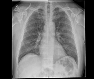
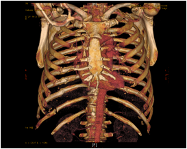
A 45 year old fit man presented 8 month after an assault with sharp localised right lumbar pain. Chest X-ray (Fig.1) and CT showed no obvious cause. A volume rendering CT scan (Fig.2); which we recommend; showed pseudoarthrosis of the right 11th rib which was successfully partially resected with an excellent result.
Figure (1) Chest X-ray showing no obvious abnormalities apart from evidence of healed 8-11th right ribs posteriorly.
Figure (2) Volume Rendering Chest CT-scan performed after ordinary CT showed no abnormalities. This clearly shows the defect in the Right 11th rib posteriorly with a picture of pseudoarthrosis.

Figure (1) Chest X-ray showing no obvious abnormalities apart from evidence of healed 8-11th right ribs posteriorly.


Figure (2) Volume Rendering Chest CT-scan performed after ordinary CT showed no abnormalities (a) Posterior view (b) Anterior view. This clearly shows the defect in the Right 11th rib posteriorly with a picture of pseudoarthrosis
First Published March 2011
Copyright Priory Lodge Education Lid. 2004 - present date
Click
on these links to visit our Journals:
Psychiatry
On-Line
Dentistry On-Line | Vet
On-Line | Chest Medicine
On-Line
GP
On-Line | Pharmacy
On-Line | Anaesthesia
On-Line | Medicine
On-Line
Family Medical
Practice On-Line
Home • Journals • Search • Rules for Authors • Submit a Paper • Sponsor us
All pages in this site copyright ©Priory Lodge Education Ltd 1994-


