Browse through our Journals...
Endocarditis in a Bengal tiger (Panthera tigris, Linnaeus, 1758).
William Pérez *, Graciela Pedrana **.
*Área de Anatomía, **Área de Histología, Facultad de Veterinaria, A. Lasplaces 1550, 11600 Montevideo, Uruguay.
Corresponding author: William Pérez, Área de Anatomía, Facultad de Veterinaria, A. Lasplaces 1550, 11600 Montevideo, Uruguay.
Summary
An 18 year old tiger from a zoo in Uruguay died of kidney failure and was submitted to necropsy. Lesions recovered in its cardiac valves were studied both macroscopically and microscopically. The valva aortae and the left atrioventricular valve were affected by endocarditis. The three semilunar valvulae of the aortic valve had proliferative lesions.
The right semilunar cusp of the aortic valve was the most affected.
Keywords:
Tiger, Panthera tigris, zoo animal, cardiac pathology, cardiac valves, wildlife disease.
Introduction
Almost all cases of endocarditis that are recognized in dogs and cats involve valve tissue, and the vast majority of the infectious agents recognized are bacterial (Kittleson and Kienle, 1998).
When bacteria colonize a heart valve, they generally produce proliferative lesions (vegetations) or destroy valve tissue. Vegetations usually result in improper coating of the valves, resulting in blood reflux. Valve destruction results in blood reflux in all instances (Kittleson and Kienle, 1998). Infectious endocarditis in feline is rarely described in the veterinary literature, with some reports of isolated cases (Shouse and Meier, 1956; Liu, 1977; De Jonghe et al., 1988; Noakes and Weisbrode, 1988; Malik et al., 1999; Kovacevic et al., 2002). In the most recent description of endocarditis in the cat (Kovacevic et al., 2002) clinical and laboratory diagnosis are reported, vegetative lesions were found in the valva aortae, but they didn't identify the causative microorganism. According to classical textbook no clinical syndrome involving the cardiovascular system has been reported in the exotic feline (Wallach and Boever, 1983). More recently the only case of endocarditis in a tiger reported in the literature was associated with the isolation of Pasteurella haemolytica (Hur et al., 2001).
We present here a new case of endocarditis in a tiger showing proliferative lesions on both the aortic valve and the left atrioventricular valve.
Materials and Methods
An eighteen year old tiger diagnosed ante-mortem with chronic renal insufficiency died in a zoo. This animal was fed exclusively on beef meat.
He had chronic renal insufficiency for the last two years, and received antibiotic treatment during the last month of his life.
Following necropsy the heart was studied isolated from the body.
The endocarditis was evident also by gross pathology and it was confirmed by histopathology. Both ventricles were cut parallel to the coronary and interventricular grooves. Both auricles were cut along their free edge. Photographs were taken with a Nikon digital camera (Fig. 1 and 2).
The Valva Aortae was fixed in 10% neutral buffered formalin for 24 hours and was processed for histological studies at the Histology laboratory: dehydrated in increasing concentrations of ethanol (70%, 95 %, 100%), immersed in chloroform for 24 hours and embedded in paraffin wax. Sections (5 mm thick) were cut using a microtome (Leica, 2135), stained with hematoxylin and eosin and Van Giesson and subjected to histological analysis. Tissue sections were mounted on positive slides.
The histological sections were photographed (Fig. 3 and 4) and images were retrieved at different magnifications with a light microscope (Olympus BX50, Tokyo, Japan) connected through a camera (SSC-C158P, Sony, Tokyo, Japan), to a personal computer containing an image analysis software, Image Pro Plus 4.0.
Results
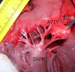
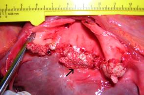
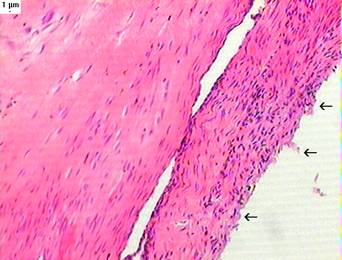
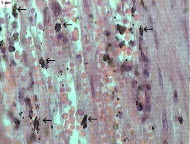
The left atrioventricular valve presented on gross examination an inflammatory reaction manifested by hyperaemia and thickening (Fig. 1). The aortic valve (Valva aortae) was affected by endocarditis, showing on all its semilunar valvulae (right, left and septal) proliferative lesions (vegetations) (Fig. 2). The vegetations vary in size and shape, but could be seen as small coalescing nodules. The right semilunar valvulae of the aortic valve were the most affected (Fig. 2, central valvulae). Around the affected valves the endocardium was minimally compromised (Fig. 1-2). On histopathology it could be seen that the vegetations consisted of bacterial colonies embedded in a connective matrix (Fig. 3-4).
Figure 1: Hyperemia and thickening of the left atrioventricular valve. AVIV: left atrioventricular valve. AV: Aortic valve. SAPM: papillary muscle.

Figure 2: Vegetative lesions of the Aortic valve. Arrow: Right semilunar valvulae of the aortic valve.

Figure 3: Aortic valve (Hematoxilin-eosin stain, 10 X). The valve shows fibrous tissue mixed with inflammatory cells and bacterial colonies (black lines and dots). Arrows indicate the damaged endocardium.

Figure 4: Aortic valve (Van Giesson stain, 40 X). Bacterial colonies in black (arrows).

Discussion and Conclusions
In the reported tiger, evident cardiac lesions affected mostly the aortic valve and the left atrio-ventricular valve (Fig 1-2). Histopathology clearly showed that these vegetations consisted of bacterial colonies embedded in a connective matrix (Fig. 3-4). It may be argued that the endocarditis caused the renal insufficiency which eventually resulted in the death of the tiger. Actually, it is acknowledged that kidney failure can be a devastating complication of infective endocarditis, occurring secondarily to the circulation of immune complexes leading to glomerulo-nephritis or as a result of renal infarction (Kittleson and Kienle, 1998). The present clinic case seems to indicate that this can be true for wild carnivores, too. In the only other report published of endocarditis in a tiger (Hur et al., 2001) there were no pictures showing the valvular lesions and no concurrent kidney failure was reported.
The authors attributed the cause of death to Pasteurella haemolytica infection. Unfortunately, in the present case no bacterial culture could be performed. The source of infection couldn't be determined either.
The morphology of cardiac vegetations can vary, depending on the infecting organism and the evolution of the disease, from small flat granular lesions to large pedunculated friable masses (Braunwald, 1992). As vegetations heal during the process of bacteriological cure, infiltration by polymorphonuclear leukocytes and fibroblasts occurs, leading to fibrosis, hyalinization, and, sometimes, calcification (Braunwald, 1992). In the reported clinic case the vegetations appeared as small coalescing nodules, suggesting that an healing process occurred, may be due to the antibiotic treatment.
Acknowledgements
We gratefully thank “Villa Dolores” Zoo and their veterinary staff for providing us with the tiger, to Elizabeth Pechiar for help in translating this paper and Dr. Alicia Baldovino.
References
1. Braunwald, E. Heart Disease. A Textbook of Cardiovascular Medicine. 4th edition. Volume 2. W. B. Saunders Company, 1992.
2. De Jonghe, S., Ducatelle, R. & Devriese, L. A. Verrucous endocarditis due to Escherichia coli in a Persian cat. In: Veterinary Record 1988; 143: 305-307.
3. Hur, K., Yoon, B. I., Yoo, H. S., Shin, N. S., Kwon, S. W., Lee, G. H & Kim, D. Y. Aortic valvular endocarditis associated with Pasteurella haemolytica in a tiger (Panthera tigris). The Veterinary Record 2001; 149: 490-491.
4. Kittleson, M. D. & Kienle, R. D. Small Animal Cardiovascular Medicine.. Mosby, 1998: 402 – 405.
5. Kovacevic, A., Allenspach, K., Kühn, N. & Lombard, C. W. Endocarditis in a cat. Journal of Veterinary Cardiology 2002; 4: 31-34.
6. Liu, S. K. Pathology of feline heart disease. In: The Veterinary Clinics of North America 1977; 7: 323-339.
7. Malik, R., Barrs, V. R., Church, D. B., Zahn, A., Allan, G. S., Martin, P., Wigney, D. I. & Love, D. N. Vegetative endocarditis in six cats. Journal of Feline Medicine and Surgery 1999; 1: 171-180.
8. Noakes M. E. & Weisbrode, S. E. What is your diagnosis? JAVMA 1988; 9: 1390-1393.
9. Shouse C. L., Meier, H. () Acute Vegetative Endocarditis in the Dog and Cat. JAVMA 1956; 129: 278-289.
10. Wallach J. D., Boever, W. J. Diseases of exotic animals: medical and surgical management. W. B. Saunders Company. Philadelphia, 1983.
© Copyright Priory Lodge Education 2007
First Published June 2007
Click
on these links to visit our Journals:
Psychiatry
On-Line
Dentistry On-Line | Vet
On-Line | Chest Medicine
On-Line
GP
On-Line | Pharmacy
On-Line | Anaesthesia
On-Line | Medicine
On-Line
Family Medical
Practice On-Line
Home • Journals • Search • Rules for Authors • Submit a Paper • Sponsor us
All pages in this site copyright ©Priory Lodge Education Ltd 1994-


